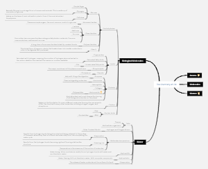
MindMap Gallery muscle tissue
Mind map of muscle tissue knowledge points, histology and embryology, muscle tissue is mainly composed of muscle cells with contractile function. There are a small amount of connective tissue, blood vessels, lymphatic vessels and nerves between muscle cells, which are used for learning and review.
Edited at 2024-04-22 08:26:10One Hundred Years of Solitude is the masterpiece of Gabriel Garcia Marquez. Reading this book begins with making sense of the characters' relationships, which are centered on the Buendía family and tells the story of the family's prosperity and decline, internal relationships and political struggles, self-mixing and rebirth over the course of a hundred years.
One Hundred Years of Solitude is the masterpiece of Gabriel Garcia Marquez. Reading this book begins with making sense of the characters' relationships, which are centered on the Buendía family and tells the story of the family's prosperity and decline, internal relationships and political struggles, self-mixing and rebirth over the course of a hundred years.
Project management is the process of applying specialized knowledge, skills, tools, and methods to project activities so that the project can achieve or exceed the set needs and expectations within the constraints of limited resources. This diagram provides a comprehensive overview of the 8 components of the project management process and can be used as a generic template for direct application.
muscle tissue
Overview
Classification
skeletal muscle
Tendons attached to bones have strong contractility, fatigue easily, and poor regeneration ability.
voluntary muscles, striated muscles
myocardium
Distributed in the heart and proximal large blood vessels, it contracts rhythmically, is not prone to fatigue, and has almost no regeneration ability.
involuntary muscles, striated muscles
smooth muscle
Distributed in visceral hollow organs and blood vessel walls, the contraction force is weak, long-lasting, and the regeneration ability is strong
involuntary muscles
constitute
Mainly composed of muscle cells with contractile function. There are a small amount of connective tissue, blood vessels, lymphatic vessels and nerves between muscle cells.
proper noun
Sarcoplasmic reticulum: smooth endoplasmic reticulum
sarcolemma: muscle cell membrane
Sarcoplasm: muscle cytoplasm
Muscle fiber: muscle cell-slender fiber type
skeletal muscle
Basic understanding
Skeletal muscles are the dynamic part of the locomotor system. Most of them are attached to bones and mainly exist in the trunk and limbs. They are controlled by human consciousness and are also called voluntary muscles.
Skeletal muscles are widely distributed in the human body, with more than 600 pieces, accounting for about 40% of the human body weight.
Each skeletal muscle has a certain position, shape, structure and auxiliary devices, and is rich in blood vessels, lymphatic vessels and nerve distribution to perform certain functions.
Divided according to location: head muscles, neck muscles, trunk muscles, diaphragm muscles, upper limb muscles, lower limb muscles, etc.
surrounded by dense connective tissue
composition
epimysium
Wrapped around the entire muscle
perimysium
The epimysium extends into the muscle, separates it to form muscle bundles, and is the connective tissue that wraps the muscle bundles.
endomysium
connective tissue outside each muscle fiber
Function
Support, connection, nutrition, functional regulation
composition
Myofibrils (subcellular structure of muscle cells) → myofibers (muscle cells, elongated fiber-shaped) → muscle bundles → skeletal muscle
skeletal muscle fibers
Light mirror structure
Cell body: long cylindrical shape
Nucleus: multinucleated cell, with dozens or even hundreds of nuclei in a muscle fiber, oval, located under the sarcolemma
Cytoplasm: filled with myofibrils arranged parallel to the long axis of the muscle fiber, in the form of filaments
periodic stripes
Horizontal stripes: The light and dark bands on each myofibril are arranged on the same plane, forming periodic horizontal stripes with alternating light and dark stripes.
Bright band: There is a dark Z line in the center. Under a polarized light microscope, the bright band appears as single refracted light, also known as the I band. Length variable.
Dark band: Under a polarized light microscope, the dark band is birefringent and anisotropic. It is also called the A band and has a constant length. Under an oil microscope or electron microscope, it can be observed that there is a more pigmented H band in the middle of the dark band, and a darker M line can be seen in the center of the H band.
Three belts and two lines: A, I, H belts; Z, M lines
Structural and functional unit of skeletal muscle fiber - sarcomere
Concept: A section of myofibril between two adjacent Z lines is a sarcomere, which is the basic structural unit of skeletal muscle contraction and relaxation.
Composition: Each sarcomere consists of 1/2 I band, A band, and 1/2 I band
The length of the dark band is constant, 1.5um, and the light band changes with the contraction and relaxation of skeletal muscles, with a maximum length of 2um.
A myofibril can be composed of hundreds of sarcomeres
Ultrastructure
myofibrils
Thick myofilament
Arranged along the long axis of myofibrils, thick myofilaments are only located in the dark band A
distributed
The center of the sarcomere, the center is fixed to the M line, and both ends are free
structure
Long balance 1.5 microns, diameter 15 nanometers
Myosin polymerizes regularly to form
Molecular structure of thick muscle filaments - myosin
Bean sprout-shaped, divided into two parts: the head and the rod. There are similar joints at the head-rod connection point, which can be flexed.
Head: towards both ends of thick myofilaments, it has ATPase activity and can bind to the actin of thin myofilaments to form a cross bridge.
Rod: Towards the middle of the thick muscle filaments, they are gathered into the main body of the thick muscle filaments with the help of the M line
Thin myofilaments
Across the light and dark bands, one end is fixed on the Z line, the other end passes through the I band of the light band, extends between the thick muscle filaments of the A band, and finally ends at the edge of the H band
distributed
One end is fixed on the Z line, the other end extends between the thick muscle filaments, and terminates on the outside of the H band.
structure
About 1 micron long and 5 nanometers in diameter
actin
Double helix chain, each monomer has a binding site for the myosin head
Tropomyosin
Double helix molecule embedded in the shallow grooves of the actin double helix chain
Troponin
Spherical, attached to tropomyosin molecules, capable of binding calcium ions
There are six thin filaments surrounding one thick filament
Band clear: only thin filaments
dark band
H-band: thick filaments only
Both sides: Thick and thin muscle filaments
Cross-section
1 thick filament-6 thin filaments
1 thin filament-3 thick filaments
transverse tubule
Concept: a tubular structure formed by the indentation of the sarcolemma into the sarcoplasm, perpendicular to the long axis of the muscle fiber, and anastomosed by the transverse tubule branches on the same plane, surrounding the myofibrils
Distribution: Located at the junction of light and dark zones
Function: conducts the excitement of the muscle membrane to the interior of the muscle fiber
sarcoplasmic reticulum
Concept: Specialized smooth endoplasmic reticulum in muscle fibers, located between the transverse tubules
Structural features
Longitudinal tubule
The part of the sarcoplasmic reticulum that surrounds myofibrils longitudinally
final pond
Flat sac formed by enlargement of both ends
triplet
Each transverse tubule is composed of terminal cisternae on both sides.
There are calcium pumps and calcium channels in the sarcoplasmic reticulum
There are calcium pumps and calcium channels on the sarcoplasmic reticulum membrane of striated muscle cells, which can pump calcium ions in the cytoplasm into the sarcoplasmic reticulum for storage against the concentration difference, making the calcium ion concentration in the sarcoplasm thousands of times higher than that in the sarcoplasm.
After the sarcoplasmic reticulum is excited, calcium channels open and stored calcium ions are released into the sarcoplasm, causing muscle cells to contract.
More mitochondria
glycogen
small amount of lipid droplets
Myoglobin (can combine with oxygen)
muscle satellite cells
Located on the surface of skeletal muscle fibers (between the sarcolemma and basement membrane), flat and protruding
After muscle fibers are injured, muscle satellite cells can proliferate and differentiate, participate in the repair of muscle fibers, and have stem cell properties.
Myofilament sliding principle
M line can shrink
A belt remains unchanged, I belt and H belt are shortened at the same time
shrink
Nerve impulses are conducted to the sarcolemma and then to the sarcoplasmic reticulum through the transverse tubules and triplets
The calcium ion channels of the sarcoplasmic reticulum open and a large amount of calcium ions enter the cytoplasm.
Calcium ions bind to troponin, causing the configuration or position of troponin and tropomyosin to change, releasing the steric hindrance effect of tropomyosin.
Actin binds to myosin heads, forming cross-bridge connections between thick and thin myofilaments
The cross bridge pulls the thin muscle filaments to slide towards the M line, shortening the season.
diastole
There is no action potential and calcium ions are pumped into the sarcoplasmic reticulum
Calcium ions are separated from troponin, and tropomyosin blocks the interaction between thick and thin myofilaments.
Thin myofilaments return, sarcomeres lengthen, and muscles relax
myocardium
Overview
Distributed on the walls of the heart and the walls of the large blood vessels adjacent to the heart
Contractions are automatic and rhythmic
muscleless satellite cells
Light mirror structural characteristics
shape
Irregular short cylindrical shape, branched and interconnected into a network
connect
Cells are connected by intercalary disks and stain darker
nucleus
1-2 nuclei, centered, oval
Cytoplasm
There are periodic striations, myofibrils are located on the periphery, and the perinuclear cytoplasm is lightly stained and contains lipofuscin.
Electron microscope structural features
Thick myofilaments, thin myofilaments and sarcomeres
The thick and thin myofilaments are not as obvious as skeletal muscle, and are separated into myofilament bundles of varying thicknesses by sarcoplasm and mitochondria.
Myofibrils vary in thickness
Mitochondria are abundant
The transverse tubule is at the level of the Z line
Ultrastructure
myocardium
The sarcoplasmic reticulum is sparse, the longitudinal tubules are underdeveloped, there are few terminal cisterns, and most of them form doublets.
Doublet: The transverse tubule is closely connected to the terminal cisterna on one side.
During contraction, calcium ions are taken in from outside the cells
leap disk
The transverse part of the intercalated disc is located at the level of the Z line. It has adhesive zonules and desmosomes that tightly connect the myocardial fibers.
There are gap junctions in the longitudinal part, which facilitate the exchange of chemical information and electrical impulses between cells and synchronize the contraction and contraction of the myocardium.
smooth muscle
distributed
Widely distributed in the walls of hollow organs such as digestive tracts, respiratory tracts, and blood vessels
Light mirror structural characteristics
Long fusiform, the size and shape vary depending on the location and the functional form of the organ, generally 200 microns long
No striations, cytoplasmic eosinophilia
Single core, rod-shaped or oval
Ultrastructure of smooth muscle fibers
Cytoplasm
There are no myofibrils, thick myofilaments, thin myofilaments and intermediate filaments can be seen. There are dense bodies in the cytoplasm. The intermediate filaments are connected between dense spots and dense bodies to form a spindle-shaped cytoskeleton.
cytoskeletal system
Dense bodies: small bodies with high electron density in the muscle plasma
Macula densa: an area of high electron density under the sarcolemma
Intermediate filaments: connected between macula densa and dense bodies, composed of desmin
membrane
There are dense spots on the membrane, and between the dense spots, the sarcolemma can be seen invading to form small depressions, which are equivalent to the transverse tubules of skeletal muscles and can transmit impulses.
Foveolae: a structure formed by the inlay of the sarcolemma
Thick and thin muscle filaments
The ratio of thick and thin myofilaments is 1:12
Thick myofilament
It is composed of myosin and is cylindrical in shape. There are cross bridges arranged in rows on the surface. Two adjacent rows of cross bridges drive in opposite directions.
Thin myofilaments
Composed of actin, one end is attached to the dense patch or dense body, and the other end is free and surrounds the thick muscle filaments
myofilament unit
Several thick and thin myofilaments gather to form a myofilament unit, also known as a contractile unit. There is only a small amount of sarcoplasmic reticulum in the cell. When the cell contracts, it also needs to absorb calcium ions from outside the cell.
sarcoplasmic reticulum
Underdeveloped, sparsely tubular
Smooth muscle fiber contraction principle
Based on the sliding theory between thick and thin myofilaments
Because the attachment points of thin myofilaments and cytoskeleton are distributed in a spiral shape, when the myofilaments slide, the muscle fibers twist in a spiral shape and their long axis shortens.
There are relatively developed gap junctions between smooth muscle fibers, which can transmit information molecules and electrical impulses, causing synchronous functional activities of adjacent muscle fibers.
Summarize
Comparison of three types of muscle tissue
distributed
Skeletal muscle: attached to bones
Myocardium: the wall of the heart and adjacent large blood vessels
Smooth muscle: mostly embedded in the walls of hollow organs
muscle type
Voluntary muscles: skeletal muscles
Involuntary muscles: cardiac muscle, smooth muscle
form
Skeletal muscles: long cylindrical, without branches (except tongue muscle)
Myocardium: short columnar with branched anastomoses
Smooth muscle: fusiform, unbranched
horizontal stripes
Skeletal muscle: obvious
Cardiac muscle: There are striations, which are not as obvious as skeletal muscle
Smooth muscle: none
nucleus
Skeletal muscle: multinucleated, located at the periphery of the cell
Cardiac muscle: 1-2 nuclei, located in the center of the cell
Smooth muscle: single nucleus, located in the center of the cell
specialized structure of sarcolemma
Skeletal muscle: transverse tubule, located at the junction of the A and I bands
Myocardium: transverse tubules, thicker, located at the Z line level
Smooth muscle: None, only sarcolemmal pits
sarcoplasmic reticulum
Skeletal muscle: developed, triplet, junction of light and dark bands
Myocardium: sparse, doublet, Z-line level
Smooth muscle: underdeveloped
gap junction
Skeletal muscle: none
Myocardium: adhesive zones, desmosomes, gap junctions, intercalary discs
Smooth muscle: gap junction
Other special structures
Skeletal muscle: sarcoplasmic reticulum
Myocardium: intercalary disc
Smooth muscle: dense bodies, dense macules