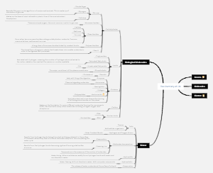
MindMap Gallery Medicine-Cardiovascular system diseases mind map
A mind map about medicine-cardiovascular system diseases, including vascular diseases, heart diseases, rheumatism, coronary heart disease, etc.
Edited at 2023-11-26 16:34:13El cáncer de pulmón es un tumor maligno que se origina en la mucosa bronquial o las glándulas de los pulmones. Es uno de los tumores malignos con mayor morbilidad y mortalidad y mayor amenaza para la salud y la vida humana.
La diabetes es una enfermedad crónica con hiperglucemia como signo principal. Es causada principalmente por una disminución en la secreción de insulina causada por una disfunción de las células de los islotes pancreáticos, o porque el cuerpo es insensible a la acción de la insulina (es decir, resistencia a la insulina), o ambas cosas. la glucosa en la sangre es ineficaz para ser utilizada y almacenada.
El sistema digestivo es uno de los nueve sistemas principales del cuerpo humano y es el principal responsable de la ingesta, digestión, absorción y excreción de los alimentos. Consta de dos partes principales: el tracto digestivo y las glándulas digestivas.
El cáncer de pulmón es un tumor maligno que se origina en la mucosa bronquial o las glándulas de los pulmones. Es uno de los tumores malignos con mayor morbilidad y mortalidad y mayor amenaza para la salud y la vida humana.
La diabetes es una enfermedad crónica con hiperglucemia como signo principal. Es causada principalmente por una disminución en la secreción de insulina causada por una disfunción de las células de los islotes pancreáticos, o porque el cuerpo es insensible a la acción de la insulina (es decir, resistencia a la insulina), o ambas cosas. la glucosa en la sangre es ineficaz para ser utilizada y almacenada.
El sistema digestivo es uno de los nueve sistemas principales del cuerpo humano y es el principal responsable de la ingesta, digestión, absorción y excreción de los alimentos. Consta de dos partes principales: el tracto digestivo y las glándulas digestivas.
cardiovascular system diseases
vascular disease
atherosclerosis
Basic lesions
secondary lesions
plaque rupture
Rupture of the fibrous cap on the plaque surface often forms atheromatous ulcers and is complicated by thrombosis. Necrotic atherosclerotic material can be discharged into the bloodstream and cause cholesterol embolism.
thrombosis
Superficial or deep intimal damage caused by plaque rupture exposes collagen in the artery wall, causing platelet aggregation to form thrombus
aneurysm formation
In severe atherosclerotic plaques, the smooth muscle of the media at the base is compressed and thinned, and its elasticity is weakened. It cannot withstand the pressure of blood flow and causes the artery wall to expand outward and form an aneurysm.
Calcification
The excessive deposition of salt and calcium in the fibrous cap and atheroma lesions causes the arterial wall to become brittle and hard, making it easy to rupture.
intraplate hemorrhage
Rupture of new capillaries within the plaque or rupture of the plaque fibrous cap
fibrous plaque
Looking under the mirror
fiber cap
The area between the subendothelium and the necrotic center is composed of SMC, dense collagen fibers, macrophages, and a small amount of elastic fibers and proteoglycans.
base
Proliferated SMC, connective tissue and inflammatory cells
lipid region
Composed of foam cells, extracellular lipids and necrotic debris
naked eye view
It is initially a gray-white plaque raised on the surface of the intima. As the collagen fibers on the surface of the plaque continue to increase and hyalinization occurs, the plaque gradually turns into a porcelain white, like a wax drop.
form
Foam cell necrosis leads to the formation of extracellular lipids and massive SMC proliferation.
atheromatous plaque
Looking under the mirror
surface
Hyalinization of collagen fibers in fibrous cap
Deep
A large amount of amorphous necrotic material, which is rich in extracellular lipids, cholesterol crystals and calcification. In severe cases, the medial SMC shrinks and becomes thinner.
form
After foam cells undergo necrosis and collapse, the lipids in their cytoplasm are released and lysosomes are released to promote necrosis and collapse of other cells.
naked eye view
surface
porcelain white
Deep
A yellow porridge-like substance formed by a mixture of lipids and necrotic disintegrating substances
Fat lines and spots
naked eye view
Fatty streaks on the aorta are commonly found on its posterior wall and branch openings, as spots 1 to 2 mm wide or yellow stripes of varying lengths.
Looking under the mirror
A large number of foam cells accumulate under the endothelial cells in fat streaks. Foam cells are round and large in size, with a large number of small vacuoles in the cytoplasm. Foam cells in fat streaks are more derived from macrophages than SMCs, and a small number of lymphocytes and neutrophils can also be seen.
Cause and pathogenesis
risk factors
Age, gender, obesity, infection
gender
Premenopausal women have higher HDL levels and lower LDL levels than men. After menopause, the difference in incidence between the sexes disappears.
age
Arterial intima gradually thicken with age
obesity
Obese people are susceptible to disease
Infect
smoking
Endothelial cell damage and increased blood carbon monoxide concentration and carboxyhemoglobin increase stimulate endothelial cells to release growth factors and promote the migration and proliferation of medial SMC into the intima.
genetics
The incidence of AS is high in patients with familial hypercholesterolemia, familial lipoprotein lipase deficiency, etc.
hypertension
Blood flow exerts strong mechanical pressure and impact on the blood vessel wall
Diseases causing secondary hyperlipidemia
Hypothyroidism and nephrotic syndrome
hyperinsulinemia
diabetes
Hyperlipidemia
Abnormally elevated plasma total cholesterol or triacylglycerols
Pathogenesis
The role of monocytes/macrophages
Monocytes adhere to the damaged endothelial surface through adhesive molecules expressed on the surface of endothelial cells, then move into the endothelial cell space and localize in the intima, transforming into macrophages. Mediated by the scavenger shoutiW, CD36 receptors and Fc receptors on their surface, macrophages engulf a large number of lipoproteins, mainly ox-LDL, and transform into macrophage-derived foam cells, which are early lesions of AS. The main components of lipopoietinosis
The role of lipids
The local accumulation of lipids in the blood vessel wall originates from plasma lipoproteins. Dyslipidemia caused by various mechanisms (TC, TAG, LDL, IDL, sLDL,) is the initiating link in the pathogenesis of AS.
Smooth muscle cell proliferation
SMC in the arterial media migrate into the intima, proliferate in the intima and synthesize EMC, transforming fat streaks into fibrous plaques and atherosclerotic plaques. Therefore, SMC proliferation is the main link involved in the formation of progressive AS lesions.
The role of endothelial cell damage
The permeability of vascular endothelial cells increases, and chronic or repeated endothelial cell damage is the initial lesion of AS.
AS lesions of vital organs
Coronary atherosclerosis
parts
Commonly seen in the right main trunk, but also in the left main trunk, left circumflex branch, and posterior descending branch. The left anterior descending coronary artery is the most serious.
Features
The lesions are often segmental and mostly occur on the heart wall side of the blood vessels. The plaques are mostly crescent-shaped, causing eccentric stenosis of the lumen. They are often accompanied by coronary artery spasm, bleeding and thrombosis.
aortic atherosclerosis
parts
Commonly found in the posterior wall of the aorta and the openings of its branches, with the abdominal aorta the most serious disease
Features
The lumen of blood vessels becomes thinner and is affected by blood pressure, which can easily form aneurysms. The rupture of aneurysms can lead to fatal bleeding.
carotid and cerebral arteries
parts
Commonly occurs in the origin of the internal carotid artery, basilar artery, middle cerebral artery, and circle of WILLIS
Features
Cerebral arteriosclerosis causes brain atrophy
mesenteric artery
Indigestion, decreased intestinal tone, constipation, abdominal pain and other symptoms
renal atherosclerosis
atherosclerotic pyknosis
Atherosclerosis of extremities
Gangrene, intermittent claudication
hypertension
concept
Hypertension refers to a sustained increase in systemic arterial blood pressure and is a clinical syndrome that can lead to changes in the heart, brain, kidneys, and blood vessels.
Types and lesions
benign hypertension
Overview
95%, long course, slow progression, more common in middle-aged and elderly people
installment
dysfunctional period
Intermittent spasm and contraction of small arteries throughout the body, no organic disease, and fluctuating elevated blood pressure
Arteriopathy stage
Arteriosclerosis
hyalinization
Arterioles spasm for a long time, plasma proteins infiltrate, SMC secretes a large amount of extracellular matrix, the wall thickens, and the lumen decreases
Arteriolar sclerosis
Involving muscular arterioles, intimal collagen fiber and elastic fiber proliferation, media SMC proliferation, blood vessel wall proliferation, and lumen stenosis
Arteriosclerosis
Significant increase in blood pressure and loss of volatility
clinical manifestations
persistently elevated blood pressure
visceral disease stage
heart
hypertensive heart disease
left ventricular hypertrophy
Centripetality
ventricular wall thickening
centrifugality
Decompensation, reduced myocardial contractility, dilation of cardiac chambers, and severe heart failure
kidney
primary granular pyknotic kidney
in general
Both kidneys are symmetrically shrunk, hard, granular in surface, thinned in cortex on cut surface, and thickened in arteriolar wall
under the mirror
Hyalinization of afferent arterioles, vitrification of diseased glomeruli, fibrosis, compensatory hypertrophy of remaining nephrons, arteriolar sclerosis, interstitial hyperplasia
brain
arteriosclerosis
Brain edema
Hypertensive crisis: sharp increase in blood pressure, spasm of small cerebral arteries, causing acute cerebral edema, symptoms of intracranial hypertension
Encephalomalacia
Multiple small lesions often occur in the putamen, caudate nucleus, optic thalamus, pons, cerebellum, cerebral cortex, etc.
cerebral hemorrhage
Often occurs in basal ganglia, internal capsule
retina
Reflects the progression of hypertension
The blood vessels are tortuous, with enhanced reflection and silver thread shape, and there are notches at the intersection of arteries and veins.
malignant hypertension
Overview
More common in young adults, blood pressure increases significantly, and the disease progresses rapidly
lesions
Hyperplastic arteriolar sclerosis, concentric layer thickening of the arterioles, necrotizing arteritis, mainly involving the kidneys
clinical
Rapid rise in blood pressure, persistent proteinuria, cerebral hemorrhage, retinal hemorrhage and detachment, heart failure
Etiology and pathogenesis
Cause
genetic and genetic factors
Hypertension has a clear hereditary tendency
Overweight obesity and abdominal obesity
High salt diet
social psychological factors
People who are mentally stressed for a long time or repeatedly may easily experience cerebral cortex dysfunction.
physical activities
It is inversely related to high blood pressure. People who lack physical activity have a higher risk of developing high blood pressure than those who are physically active.
neuroendocrine factors
Increased excitability of sympathetic nerve fibers in arterioles is the main neurological factor in the onset of hypertension
Pathogenesis
The mechanism of increased blood pressure
Sympathetic nervous system regulation
Regulation of the renin-angiotensin system
Pressor effects of epinephrine and norepinephrine
The role of vasoactive substances produced by vascular endothelium
Pathogenesis of essential hypertension
Sodium and water retention
Various reasons lead to sodium retention, which leads to water retention. The volume of plasma and extracellular fluid increases, resulting in increased cardiac output and increased blood volume.
functional vasoconstriction
There is no significant change in the structure of peripheral blood vessels. Only smooth muscle contraction reduces the caliber of blood vessels, thereby increasing peripheral resistance and causing an increase in blood pressure.
structural blood vessel wall thickening
The thickening of peripheral blood vessel walls is mainly caused by the proliferation and hypertrophy of vascular smooth muscle cells.
Aneurysm
heart disease
Rheumatism
Basic pathological changes
Rheumatic diseases of various organs
rheumatic skin lesions
rheumatic heart disease
rheumatoid arthritis
rheumatic arteritis
rheumatic encephalopathy
coronary heart disease
sudden coronary death
myocardial fibrosis
myocardial infarction
Angina pectoris
infective endocarditis
Myocarditis and cardiomyopathy
congenital heart disease
valvular heart disease
heart tumor
pericarditis