MindMap Gallery Photochemical reaction
- 6
Photochemical reaction
Substitution in photochemical reactions is a type of chemical reaction initiated by light, including radical substitution and addition-elimination substitution. When molecules absorb light energy, it is converted into chemical energy, driving the substitution reaction. These light-induced substitution reactions have specific mechanisms and pathways, leading to diverse chemical products.
Edited at 2024-12-22 09:46:55- Compound: How to use elastin
In the introduction section, elastin is a protein that plays a crucial role in the body, endowing tissues with elasticity and toughness. Its properties include high elasticity and good ductility. Elastin has a wide range of sources and is commonly found in animal connective tissues such as skin and blood vessels. When using elastin in products, the amount and method of addition should be determined based on the characteristics of the product. It has many uses and can be added to skincare products to enhance skin elasticity and reduce wrinkles; Improving food texture and enhancing taste in the food industry; In the medical field, it can be used to make elastic scaffolds such as artificial blood vessels, providing support for tissue repair. Reasonable use can play its unique value.
- Compound: Instructions for using protein
This document aims to guide the proper use of proteins. Proteins are the fundamental substances that make up life and are essential for maintaining normal physiological functions in the body, with a wide variety of types. When using protein, daily diet is an important source, such as meat, eggs, etc., which are rich in high-quality protein. Measuring protein intake can be done using professional tools or methods to ensure that the intake meets the body's needs. When mixing protein powder, it should be prepared according to the recommended ratio, and the water temperature should not be too high to avoid damaging the protein activity. Different groups of people have different protein requirements, such as athletes and fitness enthusiasts who can increase their intake appropriately to meet the needs of body repair and muscle growth.
- Compound: Usage of Unsaturated Polyester Resin
Introduction to Unsaturated Polyester Resin: It is an important thermosetting resin with various excellent properties. It has a wide range of applications and is used in the construction industry to produce fiberglass products such as doors, windows, decorative panels, etc; Used in the automotive industry for manufacturing body components. The advantages of use include simple molding process, low cost, and chemical corrosion resistance. However, it also faces challenges and limitations, such as relatively poor heat resistance and susceptibility to aging. During use, corresponding protective measures should be taken according to specific application scenarios, such as adding heat-resistant agents, antioxidants, etc., to extend their service life and fully leverage their advantages.
Photochemical reaction
- Compound: How to use elastin
In the introduction section, elastin is a protein that plays a crucial role in the body, endowing tissues with elasticity and toughness. Its properties include high elasticity and good ductility. Elastin has a wide range of sources and is commonly found in animal connective tissues such as skin and blood vessels. When using elastin in products, the amount and method of addition should be determined based on the characteristics of the product. It has many uses and can be added to skincare products to enhance skin elasticity and reduce wrinkles; Improving food texture and enhancing taste in the food industry; In the medical field, it can be used to make elastic scaffolds such as artificial blood vessels, providing support for tissue repair. Reasonable use can play its unique value.
- Compound: Instructions for using protein
This document aims to guide the proper use of proteins. Proteins are the fundamental substances that make up life and are essential for maintaining normal physiological functions in the body, with a wide variety of types. When using protein, daily diet is an important source, such as meat, eggs, etc., which are rich in high-quality protein. Measuring protein intake can be done using professional tools or methods to ensure that the intake meets the body's needs. When mixing protein powder, it should be prepared according to the recommended ratio, and the water temperature should not be too high to avoid damaging the protein activity. Different groups of people have different protein requirements, such as athletes and fitness enthusiasts who can increase their intake appropriately to meet the needs of body repair and muscle growth.
- Compound: Usage of Unsaturated Polyester Resin
Introduction to Unsaturated Polyester Resin: It is an important thermosetting resin with various excellent properties. It has a wide range of applications and is used in the construction industry to produce fiberglass products such as doors, windows, decorative panels, etc; Used in the automotive industry for manufacturing body components. The advantages of use include simple molding process, low cost, and chemical corrosion resistance. However, it also faces challenges and limitations, such as relatively poor heat resistance and susceptibility to aging. During use, corresponding protective measures should be taken according to specific application scenarios, such as adding heat-resistant agents, antioxidants, etc., to extend their service life and fully leverage their advantages.
- Recommended to you
- Outline
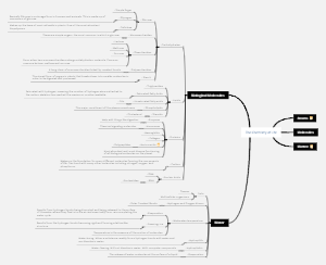
Photochemical reaction
Definition
Chemical reaction initiated by absorption of light
Involves the transformation of energy from light into chemical energy
Types of Photochemical Reactions
Primary photochemical processes
Excitation
Absorption of a photon
Electron promotion from ground state to excited state
Fluorescence
Emission of light as electron returns to lower energy level
Phosphorescence
Delayed emission of light after excitation
Internal conversion
Energy transfer without light emission
Intersystem crossing
Change in spin multiplicity during electron transition
Secondary photochemical processes
Radical reactions
Formation of radicals from excited molecules
Chain reactions involving radicals
Energy transfer reactions
Transfer of excitation energy to other molecules
Sensitized reactions
Laws Governing Photochemical Reactions
GrotthussDraper law
Only absorbed light can cause a photochemical reaction
StarkEinstein law
One molecule is activated for each photon absorbed
BunsenRoscoe law
Photochemical effect is proportional to the total light intensity and exposure time
Applications of Photochemical Reactions
Photography
Lightsensitive materials undergo changes to form images
Synthesis of chemicals
Production of pharmaceuticals, dyes, and other compounds
Environmental processes
Photodegradation of pollutants
Ozone formation and destruction
Biological systems
Photosynthesis in plants
Vision in animals
Factors Affecting Photochemical Reactions
Light intensity
Higher intensity can increase reaction rate
Wavelength of light
Specific wavelengths can trigger specific reactions
Concentration of reactants
Higher concentrations can lead to more frequent collisions and reactions
Presence of catalysts
Catalysts can alter reaction pathways and increase efficiency
Safety Considerations
Handling of lightsensitive chemicals
Proper storage and use of lightprotected containers
Eye protection
Wearing safety goggles to prevent eye damage from UV or visible light
Skin protection
Avoiding skin exposure to harmful wavelengths of light
Ventilation
Ensuring adequate ventilation to avoid inhalation of toxic vapors or gases


