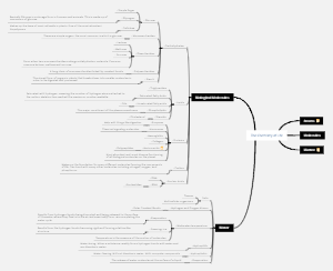
MindMap Gallery Physiology Chapter 2 Basic Functions of Cells
Regarding the mind map of Chapter 2 of Physiology, the Basic Functions of Cells, the basic functions of cells include the material transport function of the cell membrane, the bioelectrical phenomenon of the cell, the contraction function of the muscle cells, etc. These functions together maintain the normal physiological activities of the cell.
Edited at 2024-01-15 23:06:23El cáncer de pulmón es un tumor maligno que se origina en la mucosa bronquial o las glándulas de los pulmones. Es uno de los tumores malignos con mayor morbilidad y mortalidad y mayor amenaza para la salud y la vida humana.
La diabetes es una enfermedad crónica con hiperglucemia como signo principal. Es causada principalmente por una disminución en la secreción de insulina causada por una disfunción de las células de los islotes pancreáticos, o porque el cuerpo es insensible a la acción de la insulina (es decir, resistencia a la insulina), o ambas cosas. la glucosa en la sangre es ineficaz para ser utilizada y almacenada.
El sistema digestivo es uno de los nueve sistemas principales del cuerpo humano y es el principal responsable de la ingesta, digestión, absorción y excreción de los alimentos. Consta de dos partes principales: el tracto digestivo y las glándulas digestivas.
El cáncer de pulmón es un tumor maligno que se origina en la mucosa bronquial o las glándulas de los pulmones. Es uno de los tumores malignos con mayor morbilidad y mortalidad y mayor amenaza para la salud y la vida humana.
La diabetes es una enfermedad crónica con hiperglucemia como signo principal. Es causada principalmente por una disminución en la secreción de insulina causada por una disfunción de las células de los islotes pancreáticos, o porque el cuerpo es insensible a la acción de la insulina (es decir, resistencia a la insulina), o ambas cosas. la glucosa en la sangre es ineficaz para ser utilizada y almacenada.
El sistema digestivo es uno de los nueve sistemas principales del cuerpo humano y es el principal responsable de la ingesta, digestión, absorción y excreción de los alimentos. Consta de dos partes principales: el tracto digestivo y las glándulas digestivas.
Chapter 2 Basic Functions of Cells
Section 1 Material transport function of cell membrane
cell membrane structure
Dense zone - transparent zone - dense zone
basic structure of cell membrane
cell membrane: a selectively semipermeable membrane
Structure: "Liquid Mosaic Model" Hypothesis
Composition: lipids, proteins, carbohydrates
material transport across cell membranes
passive transport
Definition: A transmembrane substance transport process in which substances follow a chemical gradient or a potential gradient without consuming additional ATP chemical energy.
Diffusion power
Electrochemical potential energy (concentration difference, potential difference)
prerequisites for diffusion
The permeability of the membrane to the substance (Lipid/water solubility, molecular size, charging status, etc.)
Classification
simple diffusion
transported substances
Fat-soluble small molecule substances (O2, CO2)
facilitated diffusion
definition
The process in which some charged ions and water-soluble molecules with slightly larger molecular masses diffuse across the membrane along the concentration or potential gradient, mediated by membrane proteins.
Motive force: concentration difference, potential difference
Classification
carrier-mediated facilitated diffusion
Transported substances: nutrients
Transport mode: binding on the high concentration side → protein conformation change → dissociation on the low concentration side
Features
structure specificity
saturation phenomenon
competitive inhibition
Channel-Mediated Facilitated Diffusion
ion channel
Nature
Channel proteins with hydrophilic pores that penetrate the inside and outside of the membrane
Main features
High transport rate, ion selectivity, gating characteristics: most have valves, open and closed
Classification of gated ion channels
voltage gated channel
chemically gated channels or ligand-gated channels
Mechanically gated channel
water transport across membranes
Motive force: osmotic pressure difference (difference in concentration of water molecules)
Most cells: simple diffusion
Certain Organizations: Water Channels
Rapid transmembrane transport through special membrane proteins—aquaporins (AQP)
active transport
definition
Refers to the transport of substances across membranes against concentration or potential gradients under energy-consuming conditions.
Classification
primary active transport
Definition: Directly utilizing the energy produced by decomposing ATP
Mediated membrane proteins: ion pumps
Sodium potassium pump, calcium pump
Function: Every time a molecule of ATP is decomposed, 3 Na are pumped out and 2 K are pumped in.
Physiological significance of sodium pump
Causes high intracellular potassium ion concentration, which is necessary for many metabolic processes in the cell
Forms a concentration difference in sodium ions inside and outside the membrane to provide power for secondary active transport
Effectively maintaining the concentration difference between sodium ions and potassium ions inside and outside the cell is a prerequisite for cell bioelectric activity.
secondary active transport
Definition: indirect use of ATP energy
Required: Membrane proteins that play a coupling role - transporters
cotransport
Such as: absorption of glucose and amino acids in the epithelium of the small intestinal mucosa, reabsorption in the renal tubular epithelial cells
Antiport
Such as: myocardial sodium ion-potassium ion exchange, renal tubular sodium ion-hydrogen ion exchange
vesicle transport
The way in which macromolecules or material clumps enter and exit cells requires energy and is also an active transport.
Coming out of the cell
Definition: Refers to the process by which macromolecular substances in the cytoplasm are excreted from cells in the form of secretory vesicles.
Two forms: continuous exocytosis and intermittent exocytosis
Enter the cell
process
Contact-membrane invagination or protrusion of pseudopods-envelopment-membrane fusion and separation to form vesicles
Classification
Devour
solid matter, such as neutrophils, macrophages phagocytosis of bacteria
swallow
Liquid can be divided into liquid-phase entry into cells and receptor-mediated entry into cells.
Summarize
small molecules, ions
passive transport
Follow the electrochemical gradient and consume no energy
simple diffusion
Fat-soluble small molecule substances
Such as: O2, CO2 transmembrane transport
facilitated diffusion
Non-lipid-soluble small molecule substances that require the help of membrane proteins
Carrier mediated~
Such as: human tissue cells transport glucose, amino acids and other nutrients
channel mediated ~
Transmembrane flow of sodium and potassium ions in cellular bioelectricity
active transport
Reverse electrochemical gradient, consume energy
primary ~
Directly utilize ATP energy
sodium potassium pump
Secondary~
Small intestinal epithelial cells absorb nutrients such as glucose and amino acids
Macromolecules, lumps of matter
vesicle transport
Coming out of the cell
Nerve terminals release transmitters
Enter the cell
Neutrophils phagocytose bacteria
Chapter 3 Bioelectrical Phenomena of Cells
membrane potential
Definition: The potential difference on both sides of the cell membrane, also called transmembrane potential
Manifestations
resting potential
concept and record
definition
When a cell is in a quiet state, the potential difference between the inside and outside of the cell membrane is negative inside and positive outside.
numerical value
Take the outside of the membrane as zero potential and the negative value inside the membrane.
Different types of cells have different resting potential values.
is a stable DC potential
polarization
The inner negative and outer positive states maintained on both sides of the membrane during the resting potential
Principle of production
Base
Uneven distribution of K inside and outside the cell
The concentration difference of ions on both sides of the membrane - the driving force for ion diffusion across the membrane
There are many intracellular K and negatively charged protein macromolecules, and there are many extracellular Na and Cl-
In the quiet state, the cell membrane is mainly permeable to K
Open ion channels - conditions for ion diffusion across membranes
Resting potential and K equilibrium potential
When quiet, K channels are open, K concentration difference (power) flows out, negatively charged protein macromolecules in the membrane remain in the cell, and a potential difference between negative inside and positive outside gradually forms on both sides of the membrane. This potential difference is the resistance to K outflow. ~When the power and resistance reach a balance (the algebraic sum of the electrochemical potential energy on both sides of the membrane is zero), the net flux of K across the membrane is zero, and the membrane potential stabilizes at a certain value (K equilibrium potential)
Action potential
Concepts and Characteristics
definition
On the basis of the resting potential, excitable cells produce a rapid, short-lived, and step-expandable potential change after receiving an effective stimulus.
AP is often used as a marker of cell excitation
The Nature of Excitation: The Process that Generates Action Potentials
Expansion of the concept
Excitable cells: cells that can generate action potentials after receiving appropriate stimulation
Excitability: the ability of excitable cells to generate action potentials after being stimulated
Related terms
polarization
resting potential state
depolarization
The potential change process of decreasing negative value
Reverse polarization/overreflection
Positive inside and negative outside
repolarization
The process of depolarization and restoration of polarization
hyperpolarization
The membrane potential becomes more negative
composition
spike potential
Hallmarks of AP in neural and skeletal muscle cells
Ascending branch
descending branch
back potential
negative afterpotential
Positive back potential
feature
The “all or nothing” phenomenon
non-attenuating spread
Has a refractory period
Principle of production
Ascending branch, Na inflow, depolarization Descending branch, K outflow, repolarization
Ascending branch
Effective stimulation ~ A large number of Na channels are opened ~ Due to the Na concentration difference between high outside and low inside and the potential difference between negative inside and positive outside, Na flows inward ~ forming the action potential depolarization phase ~ during reverse polarization, the potential difference between positive inside and negative outside becomes The resistance of Na ~ When the power and resistance are balanced, the net flux of Na across the membrane is zero ~ reaching the Na equilibrium potential (overshoot vertex)
descending branch
The Na channel is closed and the K permeability increases. Due to the concentration difference and the potential difference between positive and negative inside and outside, K outflows and the membrane potential repolarizes and returns to the resting potential level.
After repolarization
Electrogenic sodium pump transport restores ion distribution inside and outside the membrane
The essential difference between action potential and resting potential
Cause and conduction of cell excitation
AP main mechanism
Effective stimulation ~ Na channel opening, large inflow of Na ~ rising branch of action potential
When the stimulation intensity is different,
Weak stimulation ~ a small amount of Na channel opening, a small amount of Na inflow ~ small degree of membrane depolarization ~ local potential
Strong stimulation ~ a large number of Na channels open, a large amount of Na inflow ~ a large degree of membrane depolarization ~ threshold potential ~ action potential
conditions that cause excitement
Parameters that measure stimulus size
stimulus intensity
duration
Intensity-time rate of change
threshold
definition
The minimum stimulus intensity that can cause tissue cells to excite under the condition that the stimulus action time and intensity-time change rate are fixed.
significance
Is a common indicator of cell excitability
Small threshold, high excitability
threshold stimulus
threshold intensity stimulus
subliminal stimulation
Stimulus with intensity less than threshold
local potential
suprathreshold stimulus
Stimulation with intensity greater than threshold
Action potential
effective stimulation
Threshold or suprathreshold stimulation that causes cells to generate action potentials
Threshold potential and action potential
Threshold potential (TP)
definition
When the stimulation intensity is increased to depolarize the membrane potential to a certain critical value, the voltage-gated Na channels on the cell membrane are rapidly activated, a large number of Na channels are opened, a large amount of Na flows in, and the rising branch of the action potential appears. This critical value is called is the threshold potential
The difference between the two is large, and the cell excitability is low.
Subthreshold stimulation and local potential
The concept of local potential
Subthreshold stimulation causes the opening of a small number of Na channels in the membrane, and potential fluctuations occur locally in the stimulated membrane.
local excitement
Depolarization reaction caused by the opening of a small amount of Na channels
Features
Not "all or nothing": the amplitude increases with the intensity of the stimulus
Attenuating step expansion: the amplitude decreases rapidly or even disappears as the distance increases
Can be superimposed on each other: no refractory period, can be summed
Meaning: Increase the excitability of cell membranes
conduction of action potential
Basic mechanism: local current theory
There is a potential difference between the excited part of the membrane and the adjacent unexcited part. The charge moves to form a local current, which depolarizes the adjacent unexcited membrane. When the threshold potential is reached, the action potential (excitation) bursts, and the entire cell membrane is excited in turn.
myelinated nerve fibers
Local current is generated at adjacent nodes of Ranvier, which is called jump conduction.
Meaning: fast conduction speed, energy saving
Periodic changes in excitability after cell excitation
cyclical change process
Absolute refractory period: excitability is 0
Relative refractory period: excitability gradually recovers
Supernormal period: excitability is higher than normal
Significance: The absolute refractory period is equivalent to the duration of the spike potential, and its length determines the maximum number of times a cell can receive stimulation and generate excitement per unit time.
contractile function of muscle cells
Contractile function of striated muscle cells
microstructure
Contains a large number of parallel arranged myofibrils and a highly developed myotube system
arranged in a highly regular and orderly manner
Myofibrils and sarcomeres
myofibrils
Bright band: variable length, the dark line in the middle is the Z line
Thin myofilament composition
Dark band: fixed length, the relatively transparent area in the middle is the H band
Thick muscle filament composition
The dark line in the center of the H band is the M line
Sarcomere
The area between every two adjacent Z lines
It is the basic structural unit of muscle cell contraction and relaxation.
Electron microscopy: regular arrangement of myofilaments
Thick myofilament
Thin myofilaments
myotubular system
membranous sac-tubular structure surrounding each myofibril
Consists of two independent piping systems
Horizontal tube (T tube)
It is formed by the inward depression of the sarcolemma, and L-type calcium channels are distributed on the membrane.
Function: Transmit electrical changes in the membrane into the interior of the cell
Longitudinal tubes (sarcoplasmic reticulum, L-tubules)
Longitudinal sarcoplasmic reticulum: membrane with calcium pump
Connecting sarcoplasmic reticulum (cisterna terminalis)
"Ca pump"
There are calcium release channels in the membrane
Three (two) conjoined
Structural basis of excitation-contraction coupling
Ca
Key factors in excitation-contraction coupling
Molecular mechanisms of striated muscle cell contraction
Myofilament sliding theory
Main content: When muscle cells contract, the shortening of myofibrils is the result of the thin myofilaments sliding toward the middle of the thick myofilaments, and the overlap of the thick and thin myofilaments.
Performance: The width of the dark band remains unchanged, but the H band and bright band become narrower. Sarcomere shortening, resulting in shortening of the overall length of myofibrils, myocytes, and muscles
Molecular composition of myofilaments
Thick myofilaments: mainly composed of myosin molecules
Thin myofilaments: composed of three protein molecules: actin, tropomyosin, and troponin
Myosin and actin are contractile proteins
Tropomyosin and troponin are regulatory proteins
muscle contraction process
Resting tropomyosin masks the cross-bridge-binding sites on actin
When the Ca2 concentration in the cytoplasm increases to 10-5M
Ca2 binds to TnC and changes the conformation of troponin
Tropomyosin allosteric, positional shift
Exposing cross-bridge-binding sites on actin
The cross bridge binds to actin and twists, causing the thin myofilaments to slide toward the M line
ATP binds to the cross-bridge, hydrolyzes ATP, and resets the cross-bridge
cross-bridge cycle, in which the muscle continuously shortens or develops constant tension
When the Ca2 concentration in the cytoplasm drops below 10-7M, Ca2 separates from troponin
Under the elastic traction of the muscle, the thin muscle filaments slide back into their original positions and the muscles relax.
Summarize
Structural basis: protein molecular composition of thick and thin myofilaments
Energy provided: ATP
The key factor that determines contraction: Ca2 concentration in the sarcoplasm
Key factors that determine the speed, degree of shortening and tension produced by a muscle: the number of cross-bridges that can participate in the cycle and the rate at which the cross-bridge circulatory activity proceeds
Excitation-contraction coupling in striated muscle cells
Concept: The intermediary mechanism of action potential in muscle cells triggering mechanical contraction
basic process
The action potential on the sarcolemma propagates along the sarcolemma and transverse canal membrane, activating L-type calcium channels on the sarcolemmal membrane and transverse canal membrane.
The calcium release channel on the terminal pool membrane is activated through changes in channel conformation (skeletal muscle) or influx of Ca2 (cardiac muscle), and Ca2 is released into the cytoplasm, increasing the cytoplasmic Ca2 concentration by more than 100 times.
Ca2 combines with troponin to initiate the sliding process of myofilaments and muscle cell contraction.
When the Ca2 concentration in the cytoplasm increases, the calcium pump on the longitudinal sarcoplasmic reticulum membrane is activated and the Ca2 in the cytoplasm is recycled into the sarcoplasmic reticulum. The Ca2 concentration in the cytoplasm decreases and the muscles relax.
Analysis of form and mechanics of striated muscle contraction (skeletal muscle)
muscle contraction pattern
Isometric and isotonic contractions
Isometric contraction
When a muscle contracts, its length remains the same and only its tension increases.
Meaning: maintain a certain posture
isotonic contraction
When a muscle contracts, its length only shortens and its tension remains unchanged.
Meaning: Complete certain physical work
Monoconstriction and tetanic contraction
single contraction
When the striated muscle is stimulated for a short time, an action potential is generated, causing the muscle to contract and relax.
The basis of tetanic contraction
The absolute refractory period is very short, so it can accept higher frequency stimulation and become excited again.
The contraction process lasts for a long time, so new stimulation can be received during the contraction process, new excitement and contraction occur, and the new contraction is the sum of the previous contraction process.
In the whole body, the outgoing impulses of the body's motor nerves are always continuous in series, and the contractions of skeletal muscles are tonic contractions.
The meaning of tetanic contraction: to produce a greater degree of tension and shortening
Mechanical analysis of muscle contraction
front load
Preload: The load a muscle bears before contracting
It determines the degree to which the muscle is stretched before contraction (i.e. the initial length), so the preload can be expressed by the initial length.
Optimum initial length: At this initial length, muscle contraction can produce maximum tension
afterload
Afterload: The load experienced by a muscle during contraction. There is always resistance to muscle shortening
Tension-speed relationship curve
P0: Isometric contraction occurs
Vmax: Send isotonic contraction
Muscle contractility
Refers to the intrinsic characteristics of muscle contraction that are independent of load.
Mainly depends on: various factors in the excitation-contraction coupling process, including L-type calcium channel activity, changes in cytoplasmic Ca2 concentration, cross-bridge function, ATPase activity, etc.