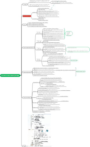
MindMap Gallery Abdominal examination
The abdomen ranges from the transverse septum to the pelvis; the abdominal body surface is bounded by the lower edges of the costal arches and xiphoid processes on both sides and the chest, and goes down to the inguinal ligaments and pubic symphysis on both sides.
Edited at 2023-11-01 20:54:03El cáncer de pulmón es un tumor maligno que se origina en la mucosa bronquial o las glándulas de los pulmones. Es uno de los tumores malignos con mayor morbilidad y mortalidad y mayor amenaza para la salud y la vida humana.
La diabetes es una enfermedad crónica con hiperglucemia como signo principal. Es causada principalmente por una disminución en la secreción de insulina causada por una disfunción de las células de los islotes pancreáticos, o porque el cuerpo es insensible a la acción de la insulina (es decir, resistencia a la insulina), o ambas cosas. la glucosa en la sangre es ineficaz para ser utilizada y almacenada.
El sistema digestivo es uno de los nueve sistemas principales del cuerpo humano y es el principal responsable de la ingesta, digestión, absorción y excreción de los alimentos. Consta de dos partes principales: el tracto digestivo y las glándulas digestivas.
El cáncer de pulmón es un tumor maligno que se origina en la mucosa bronquial o las glándulas de los pulmones. Es uno de los tumores malignos con mayor morbilidad y mortalidad y mayor amenaza para la salud y la vida humana.
La diabetes es una enfermedad crónica con hiperglucemia como signo principal. Es causada principalmente por una disminución en la secreción de insulina causada por una disfunción de las células de los islotes pancreáticos, o porque el cuerpo es insensible a la acción de la insulina (es decir, resistencia a la insulina), o ambas cosas. la glucosa en la sangre es ineficaz para ser utilizada y almacenada.
El sistema digestivo es uno de los nueve sistemas principales del cuerpo humano y es el principal responsable de la ingesta, digestión, absorción y excreción de los alimentos. Consta de dos partes principales: el tracto digestivo y las glándulas digestivas.
Abdominal examination
body surface markers
The abdomen ranges from the diaphragm to the pelvis; the abdominal body surface is bounded by the lower edges of the costal arches and xiphoid processes on both sides and the chest, and goes down to the inguinal ligaments and pubic symphysis on both sides.
1. Lower edge of the costal arch - -- composed of the costal edge formed by the connection of the 8th to 10th costal cartilages and the 11th and 12th floating ribs
2. Xiphoid process
3. Superior ventral angle - the intersection angle between the costal arches on both sides and the root of the xiphoid process
4. Umbilicus--projected backward equivalent to between the 3rd and 4th lumbar vertebrae
5. Anterior superior iliac spine
6. The outer edge of the rectus abdominis - equivalent to the continuation of the midline of the mirror bone
7. Ventral midline
8. Inguinal ligament
9.Pubic symphysis
10. Costovertebral angle - the intersection angle between the 12th rib and the spine on both sides of the back
abdominal zoning
1.Four-division method
2.Nine division method
Inspection
normal abdominal shape
In obese people (the liver is sunken) and in children (especially after meals), the front abdominal emptying is slightly higher than this level, which is called a full abdomen.
The front crotch wall of the elderly and emaciated people is slightly lower than this plane, which is called a flat abdomen.
sunken abdomen
Full abdominal depression (those who are emaciated and dehydrated)
Scaphoid abdomen-cachexia, such as tuberculosis, malignant tumors and other chronic wasting diseases
Abdominal depression during inhalation is seen in chest muscle paralysis and upper respiratory tract obstruction.
local depression
Rare
abdominal wall vein
Normal people are not exposed, they are thinner, and those with white skin are vaguely visible, but there is no tortuousness or expansion. The abdominal wall veins can be exposed in pregnancy ascites and so on.
In portal hypertension, a cluster of varicose veins can be seen radiating around, looking like a jellyfish head, and venous murmurs are often heard.
Direction of blood flow examination--acupressure method
Belly bulge
1. Full abdominal bulge
Ascites
When lying down, it looks like a frog belly, with a flat and wide appearance.
Common in cirrhosis and portal hypertension, the amount of fluid accumulated increases to the point of increased intra-abdominal pressure - umbilical protrusion
When there is inflammation or tumor infiltration in the peritoneum, the abdomen often appears sharp and convex, which is called a pointed abdomen.
Gas in the abdomen
Mostly in the gastrointestinal tract, the abdomen is spherical, the waist on both sides is not bulging significantly, and its shape does not change significantly when moving. Gas accumulation in the abdominal cavity is called pneumothorax, which is seen in gastrointestinal perforation or therapeutic artificial pneumoperitoneum.
huge intra-abdominal mass
Pregnancy, giant ovarian cysts, teratomas, etc.
2. Local bulge.
Often caused by organ enlargement, intra-abdominal tumors or inflammatory masses, gastric or intestinal flatulence, and tumors and hernias on the abdominal wall.
Differentiate between suprasabdominal wall and intra-abdominal masses
Instruct the patient to lie on his back, straighten his legs, raise his head actively, flex his neck and raise his shoulders to tense the abdominal wall muscles. If the lump becomes more obvious, it means it is located on the abdominal wall; if the lump is not obvious or disappears, it means it is located in the abdominal cavity. (Or grasp the abdominal wall) Nearly round--mostly cysts, tumors, and inflammatory masses
Those with elongated shape - mostly intestinal lesions
Those with pulsation - it may be an aneurysm, a mass on the abdominal aorta, or an organ transmitting its fluctuations
Those that shift significantly with changes in body position may be wandering organs, pedunculated masses, or masses on the omentum or mesentery; those that move with breathing are mostly subdiaphragmatic organs or masses.
Gastrointestinal and peristaltic waves
Generally invisible to normal people, it can be seen in the elderly with thin abdominal walls and sagging skin, pregnant women and extremely thin people.
The gastric peristaltic wave starts from under the left costal margin, slowly advances to the right, and disappears next to the right rectus abdominis (pyloric area). Sometimes anti-peristaltic waves can be seen.
Peristaltic waves caused by small intestinal obstruction are mostly seen in the umbilicus. In severe mechanical small intestinal obstruction, a multi-layered trapezoidal intestinal pattern can be seen in the middle of the abdomen, with undulating peristaltic waves, accompanied by high-pitched or metallic-tone bowel sounds.
Other abdominal conditions
rash
Congestive or hemorrhagic rash - seen in eruptive febrile illnesses, infectious diseases, drug allergies
Purpura, urticaria - seen in allergic diseases
Herpes on one side of the abdomen or waist (distributed along the course of the spinal nerves) - -- seen in herpes zoster
pigment
Brown pigmentation in skin folds - seen in adrenal insufficiency
Cullen's sign (blue discoloration of the skin around the umbilicus or lower abdomen) - is seen in massive intra-abdominal bleeding, ruptured ectopic pregnancy, and severe acute pancreatitis in women who are pregnant. There is brown pigmentation on the midline between the umbilicus and the pubic symphysis.
Abdominal lines
White lines
Purple lines
stretch marks
Upper abdominal pulsation
palpation
Abdominal wall tension
Increased abdominal wall tension
Reduced abdominal wall tension
Tenderness and rebound tenderness
There is no tenderness or rebound tenderness in the normal abdomen, and there is only a sense of pressure when pressed heavily. When palpating, press from shallow to deep. If pain occurs, it is called tenderness. After detecting tenderness, stop your fingers for a moment to stabilize the tenderness, and then suddenly raise your hand. At this time, if the patient feels that the abdominal pain suddenly intensifies and is accompanied by a painful expression, it is called rebound tenderness.
Tender points: 1. Gallbladder point 2. McBurney's point
liquid wave tremor
There is a large amount of free fluid in the abdominal cavity, more than 3000-4000ml, and fluctuations can be felt in the palm of the hand
Palpation of abdominal organs
Liver palpation: used to understand the location of the lower edge of the liver and the texture, edge, size, surface and pulse of the liver; use one-handed palpation or two-handed palpation
Palpation of the gallbladder: same as palpation of the liver
Palpation of the spleen: Patients with splenomegaly use one hand to palpate, but normal people use two hands to palpate.
There are three grades of splenomegaly: mild splenomegaly, moderate splenomegaly, and high splenomegaly.
Three lines: Line A and B, Line A and C, Line D and W
Palpation of the kidneys: Use both hands to palpate, noting size, texture, shape, surface condition, sensitivity, and mobility
Abdominal lumps: Feel for lumps in the abdomen and distinguish between normal and abnormal lumps
percussion
①Abdominal percussion sound: When determining the upper and lower boundaries of the liver, attention should be paid to body shape, as body shape has a certain impact on the position of the liver. The liver of an adult with a well-proportioned body is usually located on the right midclavicular line, with its upper boundary at the fifth intercostal space and its lower boundary at the lower edge of the right third rib. The distance between the two is the upper and lower diameter of the liver, which is 9 to 11cm; at the right midaxillary line On the right scapula line, its upper limit is the 7th intercostal space, and its lower limit is equivalent to the level of the 10th rib; on the right scapular line, its upper limit is the 10th intercostal space. The upper and lower borders of the liver can be one intercostal space higher in people with short and fat body types, and lower than one intercostal space in people with long and thin body types. ② Percussion of the liver and gallbladder ③ Gastric tympanic area ④ Percussion of the spleen ⑤ Percussion of the kidneys ⑥ Percussion of the bladder ⑦ Percussion of ascites: Pay attention to the differences between other diseases and ascites: 1. Intestinal obstruction 2. Those with huge ovarian cysts will also have large areas of dullness in the abdomen.
auscultation
1. Bowel sounds 2. Fluttering sounds 3. Vascular murmurs 4. Friction sounds 5. Scratching sounds
IMG_9664(20231101-190555)